- +91 7829622525
- drvikashgoyal03@gmail.com
- Apollo Sugar Clinic, 2nd floor, Golf Course Road, Road, Street Number 48, DLF Phase 1, Gurugram, Haryana 122002
- Mon - Sat 09:30 AM - 05:00 PM


EPS And RFA
EPS stands for Electrophysiology Study, while RFA stands for Radiofrequency Ablation.
An Electrophysiology Study (EPS) is a procedure used to evaluate the electrical activity within the heart to diagnose and treat abnormal heart rhythms, also known as arrhythmias. During an EPS, thin, flexible tubes called catheters are inserted into the heart through blood vessels in the groin or neck. These catheters have electrodes at their tips, which can measure electrical signals and stimulate the heart to induce arrhythmias for diagnosis. The data collected during the EPS helps cardiologists identify the specific type and location of the arrhythmia, which guides treatment decisions.
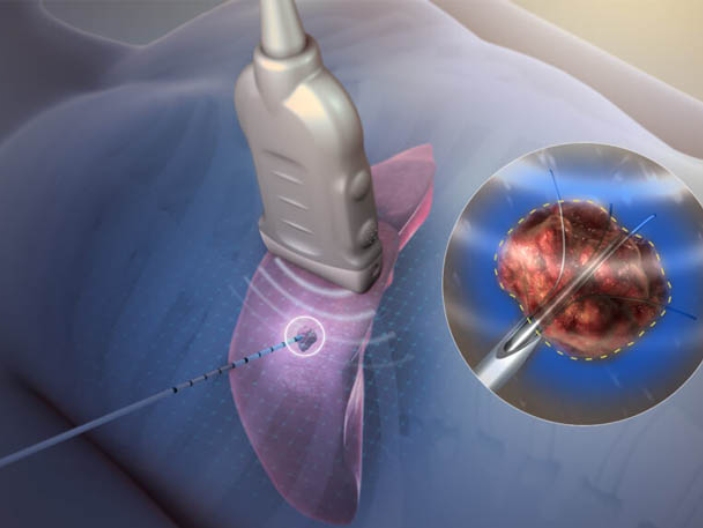
Radiofrequency Ablation (RFA) is a minimally invasive procedure often performed during an EPS to treat certain types of arrhythmias. It involves delivering high-frequency electrical energy (radiofrequency waves) through a catheter to specific areas of the heart tissue responsible for generating abnormal electrical signals. This energy heats and destroys the targeted tissue, creating scars that disrupt the abnormal electrical pathways causing the arrhythmia. By ablating or eliminating these abnormal pathways, RFA can restore normal heart rhythm and reduce or eliminate symptoms associated with arrhythmias.
Both EPS and RFA are important tools in the management of various cardiac arrhythmias, including atrial fibrillation, atrial flutter, supraventricular tachycardia, and ventricular tachycardia. These procedures are typically performed by electrophysiologists, cardiologists with specialized training in the electrical activities of the heart. They are often recommended when medications are ineffective in controlling arrhythmias or when patients experience significant symptoms or complications related to their abnormal heart rhythms.